Aphelida
This phylum includes three genera, all of which are endoparasites of algae.
Zopf described Aphelidium in 1885 for an endoparasite of the green algae Coleochaete, A. deformans, the type species. The genus is characterized by the production of posteriorly uniflagellate zoospores (Karpov et al. 2014a).
Scherffel erected the genus Amoeboaphelidium in 1925 for a similar endoparasite of the diatom Achananthes, and Am. achnanthides is the type species for the genus. The motile cells of Amoeboaphelidium are amoeboid and produce filose pseudopodia and no flagellae, distinguishing them from motile cells of Aphelidium. However, recently a pseudocilium structure has been discovered among some members of this genus (Karpov et al. 2013, Letcher et al. 2015).
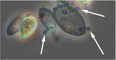
The photomicrograph at the left shows encysted motile cells of Amoeboaphelidium protococcarum at the surface of Scenedesmus dimorphus cells. The white arrows point out the encysted cells with penetration tubes entering the host cell. It is through these penetration tubes that the parasite ejects its protoplast into its host to become a multinucleate plasmodium, which replaces host cytoplasm (Letcher et al. 2015).
Schweikert and Schnepf described Pseudaphelidium in 1996 for an endoparasite of marine diatoms, with P. drebesii as the type species. Motile cells of this genus are posteriorly uniflagellate. Unlike the other two genera, Pseudaphelidium everts a tube from the encysted motile cell, which squeezes through gaps in frustules of the diatom host. Through this tube the parasite then discharges its protoplast into the host cell (Schweikert and Schnepf 1997). The parasite's plasmodium phagocytizes host cytoplasm and develops a large digestive vacuole. At maturity, the unwalled plasmodium fills the host as a hollow sphere (Schweikert and Schnepf 1996). Rather than releasing zoospores directly, it discharges amoeboid cells, which encyst, and then release zoospores.
Most recently Karpov et al. (2016) described the new genus Paraphelidium with amoeboflagellate zoospores. Molecular phylogenetic analysis places the genus in a lineage among environmentally derived phylotypes.
- Aphelidium
- Amoeboaphelidium
- Paraphelidium
- Pseudaphelidium
Zopf described Aphelidium in 1885 for an endoparasite of the green algae Coleochaete, A. deformans, the type species. The genus is characterized by the production of posteriorly uniflagellate zoospores (Karpov et al. 2014a).
Scherffel erected the genus Amoeboaphelidium in 1925 for a similar endoparasite of the diatom Achananthes, and Am. achnanthides is the type species for the genus. The motile cells of Amoeboaphelidium are amoeboid and produce filose pseudopodia and no flagellae, distinguishing them from motile cells of Aphelidium. However, recently a pseudocilium structure has been discovered among some members of this genus (Karpov et al. 2013, Letcher et al. 2015).
The photomicrograph at the left shows encysted motile cells of Amoeboaphelidium protococcarum at the surface of Scenedesmus dimorphus cells. The white arrows point out the encysted cells with penetration tubes entering the host cell. It is through these penetration tubes that the parasite ejects its protoplast into its host to become a multinucleate plasmodium, which replaces host cytoplasm (Letcher et al. 2015).
Schweikert and Schnepf described Pseudaphelidium in 1996 for an endoparasite of marine diatoms, with P. drebesii as the type species. Motile cells of this genus are posteriorly uniflagellate. Unlike the other two genera, Pseudaphelidium everts a tube from the encysted motile cell, which squeezes through gaps in frustules of the diatom host. Through this tube the parasite then discharges its protoplast into the host cell (Schweikert and Schnepf 1997). The parasite's plasmodium phagocytizes host cytoplasm and develops a large digestive vacuole. At maturity, the unwalled plasmodium fills the host as a hollow sphere (Schweikert and Schnepf 1996). Rather than releasing zoospores directly, it discharges amoeboid cells, which encyst, and then release zoospores.
Most recently Karpov et al. (2016) described the new genus Paraphelidium with amoeboflagellate zoospores. Molecular phylogenetic analysis places the genus in a lineage among environmentally derived phylotypes.
Life history of Aphelida
Gromov (2000) and Karpov et al. (2014b) have summarized the Phylum Aphelida and discussed life histories of these organisms. Motile cells of the parasite move to the host cell where they attach and encyst. The encysted cell penetrates the host wall with a tube and ejects its uninucleate protoplast into the host. The protoplast phagocytizes host cytoplasm as it grows into a multinucleate plasmodium, eventually replacing host cytoplasm and filling up the wall of the host. The plasmodium than cleaves into motile spores, which escape and continue the life cycle on a fresh host. Production of round to oval resting spores is reported in some species.
References
Gromov 2000. Entomological Review 80(Suppl 1): 26–34; translated from Zool Zurn 79:517–525 (2000).
Karpov et al. 2013. Protist 164: 195-205.
Karpov et al. 2014a. Protist 165: 512-526.
Karpov et al. 2014b. Frontiers in Microbiology 5: Article 112.
Karpov et al. 2016. Journal of Eukaryotic Microbiology doi 10.1111/jeu.12352-4854
Letcher et al. 2015. Mycologia 107: 522-531.
Scherffel 1925. Archiv der Protistenkund 52: 1-141.
Schweikert and Schnepf 1996. Archiv der Protistenkund 147: 11-17.
Schweikert and Schnepf 1997. Protoplasma 199: 113-123.
Zopf 1885. Zur Morphologie und Biologie der niederen Pilztziere (Monadinen).